 |
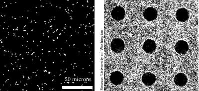
| Description: The left image shows microtubule molecules that contain fluorescent dye spreading out across a surface coated with the protein kinesin. The image on the right, taken 40 minutes later, shows the micro- tubules spread out to reveal the surface topology. |
| Source: University of Washington |
| Story: Cell
parts paint picture TRN July 10/17, 2002 |
| TRN Categories: Biotechnology; Materials Science and Engineering; Data Acquisition |
| Form: Still |
|
TRN
Newswire and Headline Feeds for Web sites
|
© Copyright Technology Research News, LLC 2000-2008. All rights reserved.