Surgeons
gain ultrasonic vision
By
Eric Smalley,
Technology Research NewsSurgeons often use ultrasound to see where they're going as they poke around their patients' insides with needles and tubes during surgery.
The problem is they have to shift their attention between the patient and the ultrasound monitor, making it difficult to coordinate what their eyes are seeing with what their hands are doing.
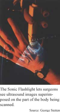
Researchers from University of Pittsburgh and Carnegie Mellon University have developed a device that overlaps the two views, giving surgeons the illusion of peering directly into their patients' bodies.
"We aim to provide a natural, unified environment for hand-eye coordination during invasive procedures guided by tomographic images," said George Stetten, an assistant professor of bioengineering at the University of Pittsburgh and a research scientist at the Robotics Institute at Carnegie Mellon University.
The handheld real-time tomographic reflection device, or Sonic Flashlight, keeps the ultrasound image aligned with the patient even when the surgeon shifts her point of view. "The Sonic Flashlight uses a flat-panel monitor and a half-silvered mirror to produce a virtual image of the ultrasound slice at its actual location within the body," said Stetten.
The device has three components: an ultrasound monitor, a half-silvered mirror, which reflects a portion of the light that hits it and lets the remainder pass through, and an ultrasound transducer, which generates the sound waves and records their reflections. The device looks like a rectangular box with a pistol grip at one end and a windshield -- the half silvered mirror -- on top of the box. A small monitor is also mounted on top of the box at an angle to the mirror. The mirror and monitor form a V-shape when viewed from the side.
The surgeon presses the transducer against the patient's body and looks through the half-silvered mirror at the patient while the ultrasound monitor projects a view of the patient's insides onto the mirror. The real and virtual views line up because each point on the ultrasound display and its corresponding point in the patient are on opposite sides of the mirror at precisely the same angle, said Stetten.
The key to keeping the views aligned is that the device's components are fixed to each other so moving the transducer also changes the position of the ultrasound monitor and half-silvered mirror. This keeps the image of the sound waves and the parts of the patient they are depicting at the correct angles on opposite sides of the mirror.
Other approaches to merging virtual and real views of patients use head-mounted displays and tracking devices to keep the views aligned, but these can be cumbersome and have limited fields of view.
"Although there have been other systems to register medical image data with the surgeon's perspective, nice aspects of this approach include the real-time registration and the optical illusion of the image appearing beneath the skin," said T. Douglas Mast, an assistant professor of acoustics at Pennsylvania State University.
The device needs to be carefully calibrated and tested before it can be considered for surgical use, said Mast. "That calibration will actually be extremely difficult, since the inhomogenous sound speed in the body must be accounted for. This is a big impediment toward surgical use, since surgeons need very accurate indications of where their [instruments] are going," he said.
It is likely to take between two and five years before practical applications are possible, said Stetten.
Stetten's research colleagues were Vikram Chib, Daniel Hildebrand and Jeanette Bursee of the University of Pittsburgh. They presented the research at the IEEE/ACM International Symposium on Augmented Reality in New York in October. The research was funded by the Whitaker Foundation and Carnegie Mellon University.
Timeline: 2-5 years
Funding: Private; University
TRN Categories: Data Representation and Simulation; Applied Technology
Story Type: News
Related Elements: Technical paper, "Real Time Tomographic Reflection: Phantoms for Calibration and Biopsy," IEEE/ACM International Symposium on Augmented Reality, New York, October, 2001
Advertisements:
December 19, 2001
Page One
LED fires one photon at a time
Chips turn more heat to power
Data protected on unlocked Web sites
Surgeons gain ultrasonic vision
Temperature changes laser color

News:
Research News Roundup
Research Watch blog
Features:
View from the High Ground Q&A
How It Works
RSS Feeds:
News
Ad links:
Buy an ad link
| Advertisements:
|
 |
Ad links: Clear History
Buy an ad link
|
TRN
Newswire and Headline Feeds for Web sites
|
© Copyright Technology Research News, LLC 2000-2006. All rights reserved.