Implant
links nerve cells to electronics
By
Kimberly Patch,
Technology Research NewsAlthough nerve cells and electronics work quite differently, scientists have for decades been trying to connect them.
Researchers from the University of Michigan have improved the connection by coating electrodes with a carefully controlled mix of plastic and protein.
The work is a step toward achieving finer control of prosthetic limbs, restoring damaged senses like sight and hearing, and providing direct connections between the brain and equipment like cameras.
Electronics and neurons both communicate using electrical pulses, but the physics involved in each is different. Electronics use currents of electrons, which carry a negative charge, while neurons use ions, which have an imbalance of electrons, and so can carry a negative or positive charge.
Electronics generate pulses by using energy to move electrons along metal wires. A nerve cell generates a pulse from one end of its oblong shape to the other by using energy to pump positively-charged sodium, potassium, or calcium ions across the membrane at one end, then allowing the ions to flow back all at once. Nerve cells range in length from 10 microns to 1 meter. A micron is one millionth of a meter.
The researchers' electrode coating is a mix of a polymer that conducts electrical current and proteins that allow similar interactions with ions. The polymer also contains a substance that encourages neurons to grow around the metal electrode.
The key to the method is that the coating is not smooth, but has a fuzzy surface that increases the contact area between electrode and brain tissue, said David C. Martin, an associate professor of materials science and engineering, and biomedical engineering at the University of Michigan. "A large surface area can be packed into a small volume if the structure is fuzzy," he said.
The fuzzy structure "makes it possible to accommodate the dramatic difference in mechanical properties between the soft brain tissue and the hard silicon devices," Martin said. More surface area provides more places where neurons can bind to the electrode, gives neurons better access to the growth substance contained in the polymer, and makes it easier for electric charge to move across the interface, he said.
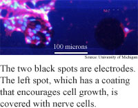
To test their method, the researchers coated electrodes a few millimeters long, 100 microns wide and 15 microns deep, and encouraged rat glial cells to grow on the devices. Glial cells provide support for neurons, which conduct the impulses of biological communications systems. A micron is one thousandth of a millimeter, and a human hair is about 75 microns in diameter. They also implanted the probes into living guinea pigs.
The fuzzy surface allowed electric current to flow more easily at the 1-kilohertz frequency that corresponds to the 1-millisecond width, or wavelength, of a neural pulse. The surface decreased the impedance, or resistance to electron flow by one or two orders of magnitude, according to Martin. "The lower the impedance at 1 kilohertz, the easier it is for electrical information to transfer from the probe to cells or vice versa," he said.
As the coating gets thicker, its surface becomes more and more rough, increasing the surface area, but there's also a point at which it becomes too thick and begins to increase resistance, said Martin. "There is an optimal film thickness at which the [charge] transport is the easiest," he said.
The experiments also showed that the coatings encouraged neurons to bind to the electrodes. "We see neural cells attached to the surface of the probe after it is removed, whereas uncoated probes come out clean," said Martin.
The researchers are currently working on coating electrodes with softer films of hydrogel materials that swell when they come in contact with water, and could increase the surface area further, according to Martin. They are also preparing to do tests with implanted probes in in rats.
The coatings could eventually be used for other types of devices, like pacemakers, that interface electronically with living tissue, Martin said.
Eventually the technique could be used to make new connections between electronics and tissue to restore sight or hearing, said Martin. "Another possible outcome may be the ability to control robotic equipment, or prosthetic... limbs."
This type of interface could also allow for direct brain interfaces to cameras that detect things humans cannot, like infrared light or magnetic fields, Martin said.
Martin's research colleagues were Yinghong Xiao, Junyan Yang, David Lin, Donghwan Kim, and Xinyan Cui. They presented the research at the 34th central regional meeting of the American Chemical Society on June 27 at Eastern Michigan University. The research was funded by the the National Institutes of Health (NIH) and the National Science Foundation (NSF).
Timeline: Unknown
Funding: Government
TRN Categories: Biotechnology; Materials Science and Engineering; Human-Computer Interaction
Story Type: News
Related Elements: Technical paper, "Surface Modification of Neural Recording Electrodes with Conducting Polymer/biomolecule Blends," Journal of Biomedical Materials Research, August, 2001 .
Advertisements:
July 24/31, 2002
Page One
Disks set to go ballistic
Two-step queries bridge search and speech
Implant links nerve cells to electronics
Silicon chips set to go atomic
Light switch promises powerful computers
News:
Research News Roundup
Research Watch blog
Features:
View from the High Ground Q&A
How It Works
RSS Feeds:
News
Ad links:
Buy an ad link
| Advertisements:
|
 |
Ad links: Clear History
Buy an ad link
|
TRN
Newswire and Headline Feeds for Web sites
|
© Copyright Technology Research News, LLC 2000-2006. All rights reserved.